Ultrasonic Color Doppler Diagnostic Systems: Best Techniques and Protocols 2026
What is it used for in 2026
Ultrasonic Color Doppler Diagnostic Systems have become essential tools in various medical applications in 2026. They are primarily used for imaging and assessing blood flow and cardiac activity. Laboratory professionals utilize these systems for diagnostic purposes in obstetrics, gynecology, urology, cardiology, and vascular assessments. The advanced imaging capabilities help in evaluating the heart's structure and function, detecting abnormalities, and guiding treatment decisions.
History and evolution of the technology
The evolution of Doppler ultrasound technology began in the 1950s when the principles of ultrasound were first applied in medical diagnostics. Initially limited to basic imaging, advancements have led to the development of color Doppler systems capable of providing real-time visualization of blood flow. Over the decades, these systems have integrated advanced software algorithms, enhanced imaging techniques, and user-friendly interfaces, making them indispensable in modern diagnostics.
How to use it step by step
Using an Ultrasonic Color Doppler Diagnostic System involves several steps:
- Preparation: Ensure the equipment is calibrated and ready for use. Check for proper probe function and connection.
- Patient Preparation: Explain the procedure to the patient and position them comfortably. Apply a suitable coupling gel to enhance sound transmission.
- Select the Mode: Choose the appropriate imaging mode based on the diagnostic requirements (e.g., 2D, Doppler).
- Perform Imaging: Move the probe over the area of interest, adjusting angles and positions to capture optimal images.
- Analyze Results: Review the captured images and Doppler waveforms, interpreting them based on established criteria.
- Documentation: Save the images and generate reports as needed, ensuring compliance with data management protocols.
Best techniques and protocols
To achieve the best results with Ultrasonic Color Doppler Diagnostic Systems, adhere to the following techniques and protocols:
- Regularly calibrate and maintain the equipment to ensure accuracy.
- Use appropriate gel for better sound conduction and avoid artifacts.
- Utilize standardized protocols for positioning the probe for different diagnostic purposes.
- Stay updated with the latest imaging techniques and software enhancements to improve diagnostic accuracy.
- Engage in continuous education to become proficient in interpreting Doppler waveforms and assessing vascular conditions.
Practical applications by laboratory type
Ultrasonic Color Doppler Diagnostic Systems find practical applications across various laboratory settings:
- Cardiology Labs: Assess cardiac function, evaluate valvular and congenital heart diseases.
- Obstetric Clinics: Monitor fetal development and placental blood flow.
- Vascular Clinics: Diagnose vascular occlusions, stenosis, and thrombosis.
- Urology Departments: Evaluate kidney blood flow and detect renal abnormalities.
Regulations, standards and certifications
In 2026, the use of Ultrasonic Color Doppler Diagnostic Systems is governed by various regulations and standards, including:
- ISO and IEC standards for medical electrical equipment.
- FDA regulations for safety and effectiveness of ultrasound devices.
- Compliance with HIPAA for patient data privacy and security.
- Certification from relevant health authorities to ensure quality and reliability.
Comparison with alternative technologies
While Ultrasonic Color Doppler Diagnostic Systems are widely used, other technologies such as MRI and CT scans offer different advantages:
- Ultrasound: Non-invasive, real-time imaging, and cost-effective.
- MRI: Superior soft tissue contrast but more expensive and time-consuming.
- CT Scans: Excellent for detailed cross-sectional imaging but involves radiation exposure.
Comparison of available models
| Model | Best for | Key specs | Recommended use case |
|---|---|---|---|
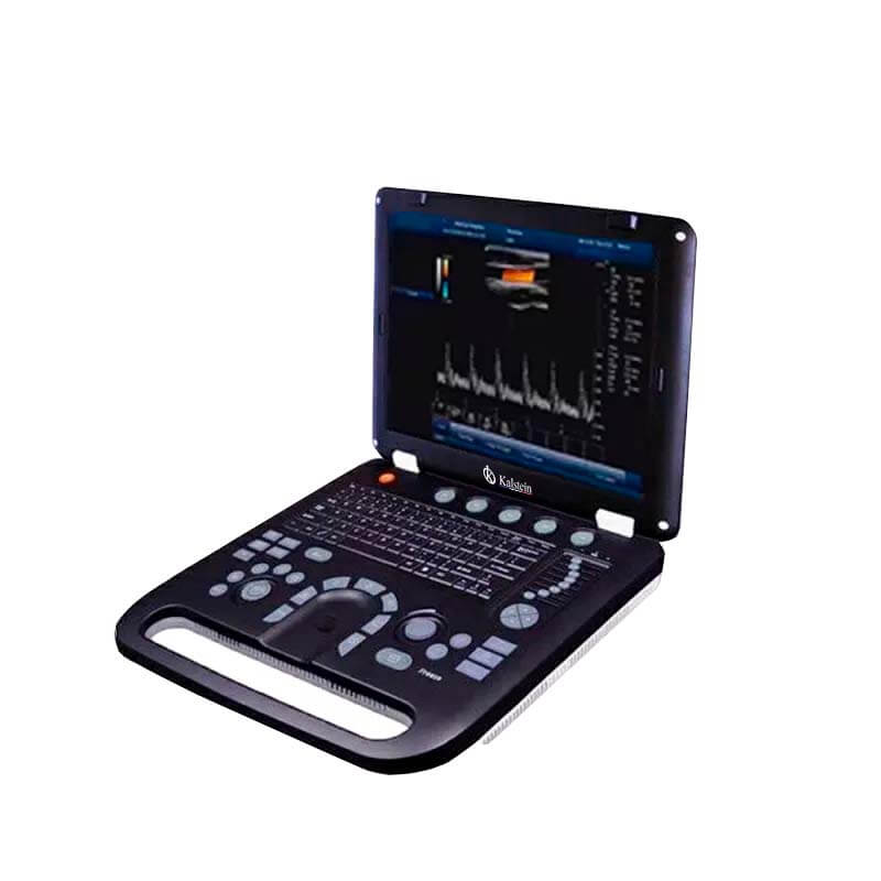
| YR05148 | General ultrasound with color Doppler | 15" LED screen, dual and triplex modes, automatic measurements | Routine assessments in various specialties |
| YR05149 | Mobile ultrasound applications | 15" LED screen, multiple USB ports, DICOM compatibility | Dynamic clinical environments requiring mobility |
| YR05150 | Portable Doppler diagnostics | 15" color screen, built-in battery, multi-mode imaging | Emergency medicine and outpatient settings |
| YR05151 | High-quality vascular assessments | 15" color screen, advanced imaging modes, PC-based | Specialized vascular labs |
| YR05152 | Cardiac evaluations | 15" screen, CW function, advanced imaging capabilities | Cardiology departments |
| YR05153 | General diagnostics with portability | 15" LED screen, multiple modes, adjustable print area | Multi-specialty use in various locations |
Common mistakes and how to avoid them
Laboratory professionals should be aware of common mistakes when using Ultrasonic Color Doppler Diagnostic Systems:
- Improper gel application: Ensure sufficient gel is used to avoid air pockets.
- Poor probe positioning: Familiarize yourself with optimal angles for different imaging requirements.
- Neglecting calibration: Regularly check and calibrate systems for accurate readings.
- Inadequate patient preparation: Communicate effectively with patients to minimize movement during scans.
Maintenance, calibration and good practices 2026
Maintaining Ultrasonic Color Doppler Diagnostic Systems is vital for their longevity and performance:
- Perform routine cleaning of probes and equipment to prevent contamination.
- Calibrate systems monthly or as recommended, ensuring accuracy.
- Keep software updated to access new features and improvements.
- Train staff regularly on best practices and equipment handling.
Cost-benefit analysis 2026
When considering Ultrasonic Color Doppler Diagnostic Systems, evaluate the following cost-benefit aspects:
- Initial investment: Compare prices of different models and their features.
- Operational costs: Account for maintenance, consumables, and training.
- Return on investment: Assess how enhanced diagnostic capabilities can lead to improved patient outcomes and increased referrals.
Frequently asked questions
What should I consider when choosing an Ultrasonic Color Doppler system?
When selecting a system, consider the imaging capabilities, portability, user interface, and compatibility with existing laboratory workflows.
How do I ensure accurate Doppler measurements?
To achieve accurate measurements, ensure proper probe positioning, adequate gel application, and system calibration before use.
What are the benefits of using color Doppler ultrasound technology?
Color Doppler technology provides real-time blood flow assessment, non-invasive imaging, and enhanced diagnostic accuracy for various medical conditions.
How can I improve my skills in using Ultrasonic Color Doppler systems?
Engage in continuous education, attend workshops, and practice regularly with supervision to enhance your skills and confidence in using these systems.
What maintenance steps should I perform on a Doppler ultrasound system?
Regularly clean the equipment, calibrate systems as per guidelines, and ensure software is updated to maintain optimal performance and reliability.
Are there any specific certifications required for operating these systems?
Yes, operators may need to adhere to certifications related to medical imaging, safety standards, and patient privacy regulations.
If you are looking for a fusion of innovation and quality, you have come to the right place. At Kalstein, we offer you the luxury of exploring our exclusive catalog of laboratory equipment. We manufacture every device to the highest standards of excellence. Our intuitive and seamless online purchasing channels are designed for your convenience, securing the most competitive prices. Hesitate no longer — we bring science to life, it is time to become part of our community.